Home

pavimento incondizionato cabina electron microscope eye Marco Polo Schiacciare Collettore

The Human Eye and Adaptive Optics | IntechOpen

up close + very personal | Human eye, Eye anatomy, Microscopic photography

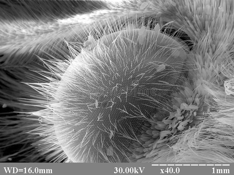
Silicon Balls On Moths Eye Imaged In A Scanning Electron Microscope  High-Res Stock Photo - Getty Images
Silicon Balls On Moths Eye Imaged In A Scanning Electron Microscope High-Res Stock Photo - Getty Images

File:Inner Structure of the Fly's Eye on scanning electron microscope.jpg -  Wikimedia Commons
File:Inner Structure of the Fly's Eye on scanning electron microscope.jpg - Wikimedia Commons

21 Extreme Close Ups of the Human Eye » TwistedSifter

electron microscopy | The Dragonfly Woman

PDF] Scanning electron microscopic studies of the zonular apparatus in  human and monkey eyes. | Semantic Scholar
PDF] Scanning electron microscopic studies of the zonular apparatus in human and monkey eyes. | Semantic Scholar

NZ Nano: Scanning electron microscope image of a cats eye

বাঘযাত্রা ।। Journey with Tigers - 👁 You have beautiful eyes! Really?  Microscopic, Macroscopic and Coloured Scanning Electron micrograph (SEM) of  rods (blue) and cones (purple) of human eye. 📸 #Google | Facebook
বাঘযাত্রা ।। Journey with Tigers - 👁 You have beautiful eyes! Really? Microscopic, Macroscopic and Coloured Scanning Electron micrograph (SEM) of rods (blue) and cones (purple) of human eye. 📸 #Google | Facebook
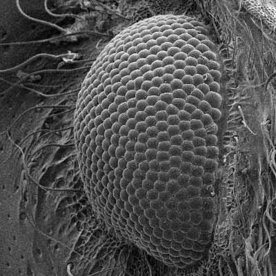
WMG on Twitter: "I spy a bug's eye! Pop in and see us at the  #WarwickFamilyDay on Sunday Take a closer look at bugs and insects using  our powerful desktop scanning electron
WMG on Twitter: "I spy a bug's eye! Pop in and see us at the #WarwickFamilyDay on Sunday Take a closer look at bugs and insects using our powerful desktop scanning electron
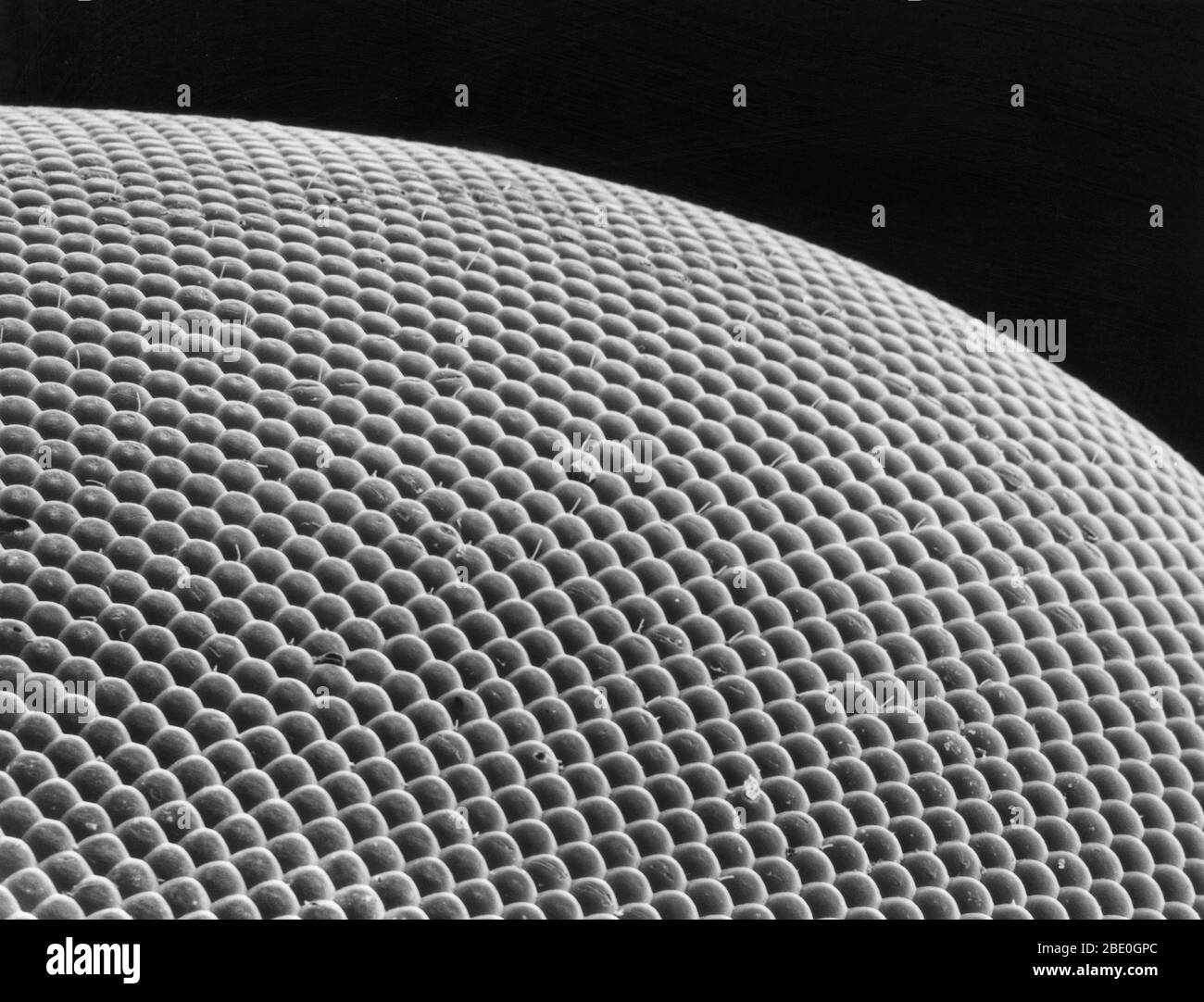
Deer fly eye viewed under a scanning electron microscope. The large, green  iridescent eyes of the deer fly are made up of thousands of individual  lenses which allow the fly to see
Deer fly eye viewed under a scanning electron microscope. The large, green iridescent eyes of the deer fly are made up of thousands of individual lenses which allow the fly to see
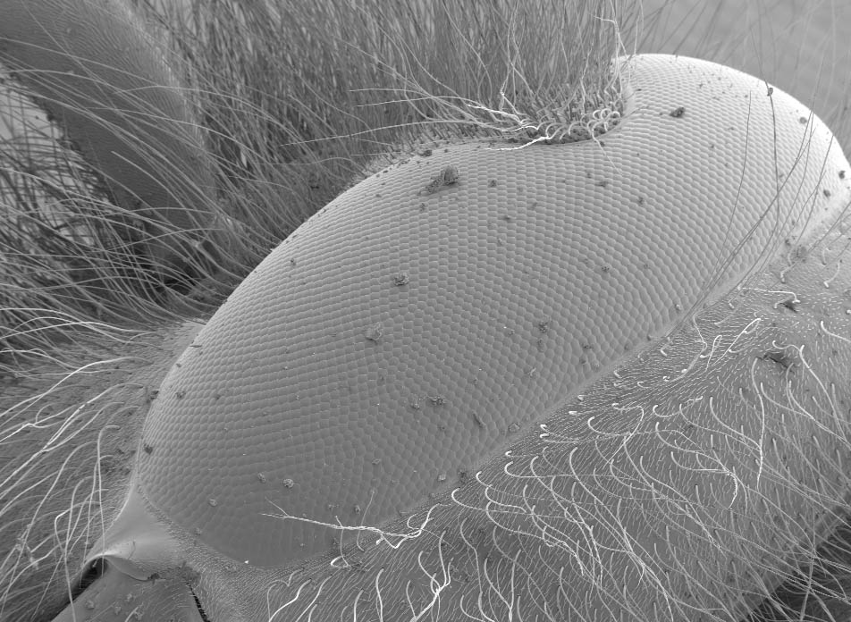
Butterfly Eye Structure - 1 Stock Image - Image of biology, macroworld:  183780953
Butterfly Eye Structure - 1 Stock Image - Image of biology, macroworld: 183780953

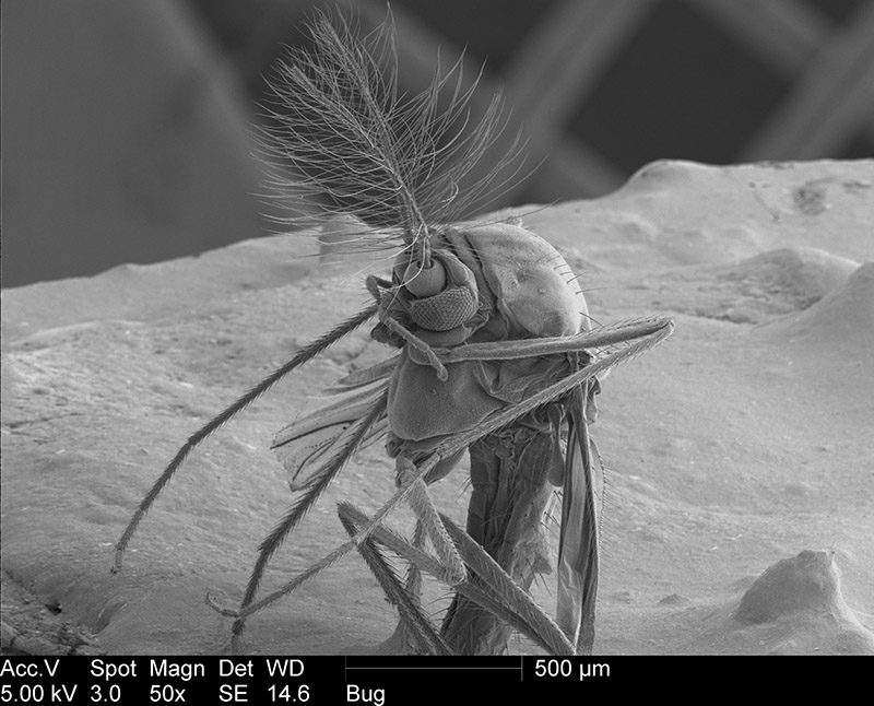
10 Fascinating Photos Of A Gnat Under An Electron Microscope. The Eye Is  Amazing
10 Fascinating Photos Of A Gnat Under An Electron Microscope. The Eye Is Amazing

The Better to See Things With | Virginia Wesleyan University

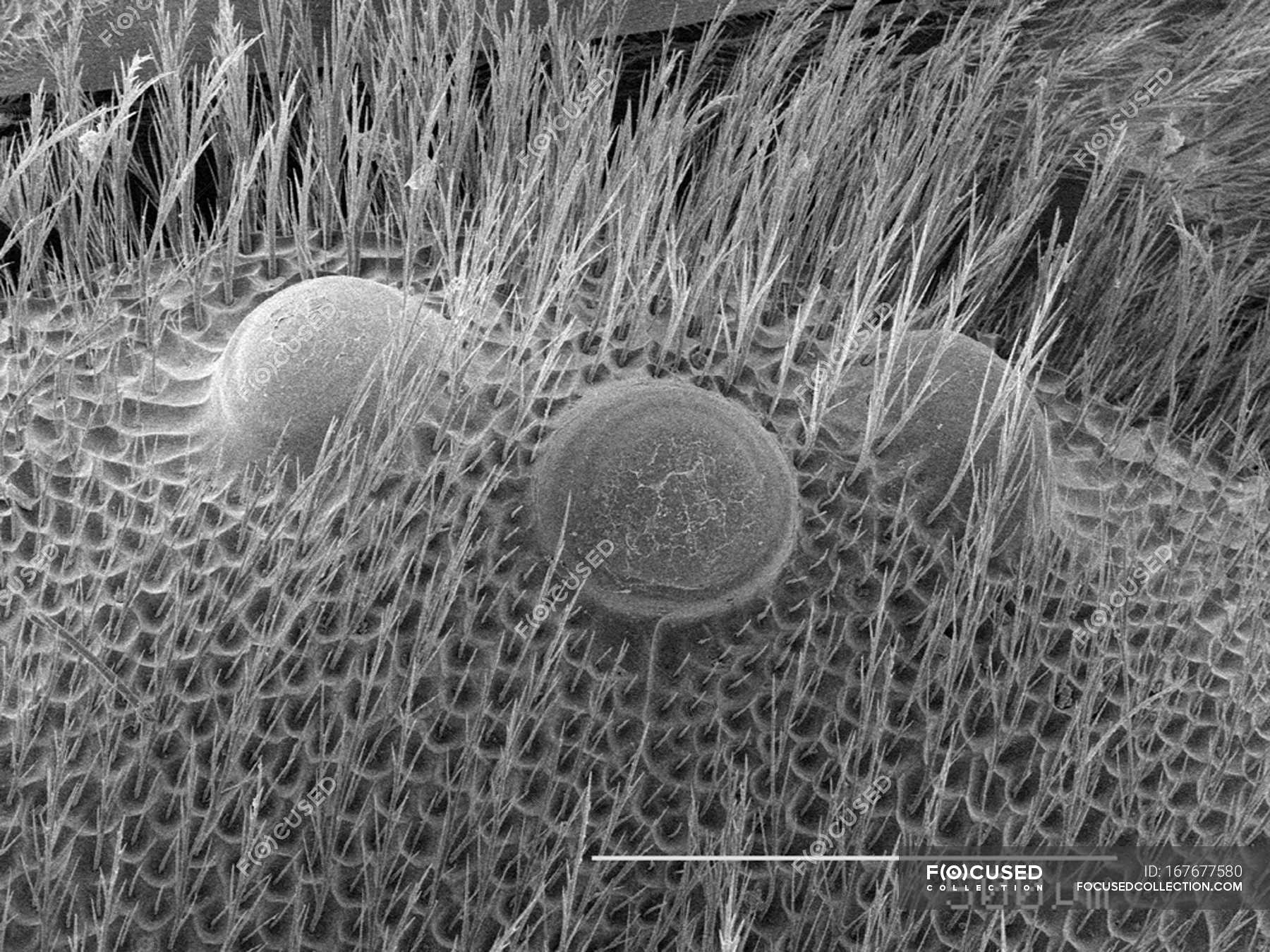
Scanning electron micrograph of eyes of bee — laboratory, microscope -  Stock Photo | #167677580
Scanning electron micrograph of eyes of bee — laboratory, microscope - Stock Photo | #167677580

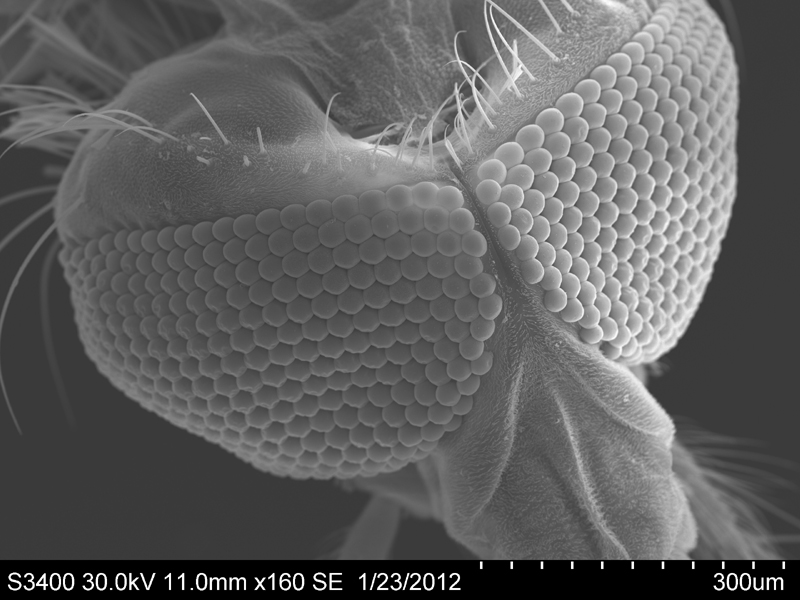
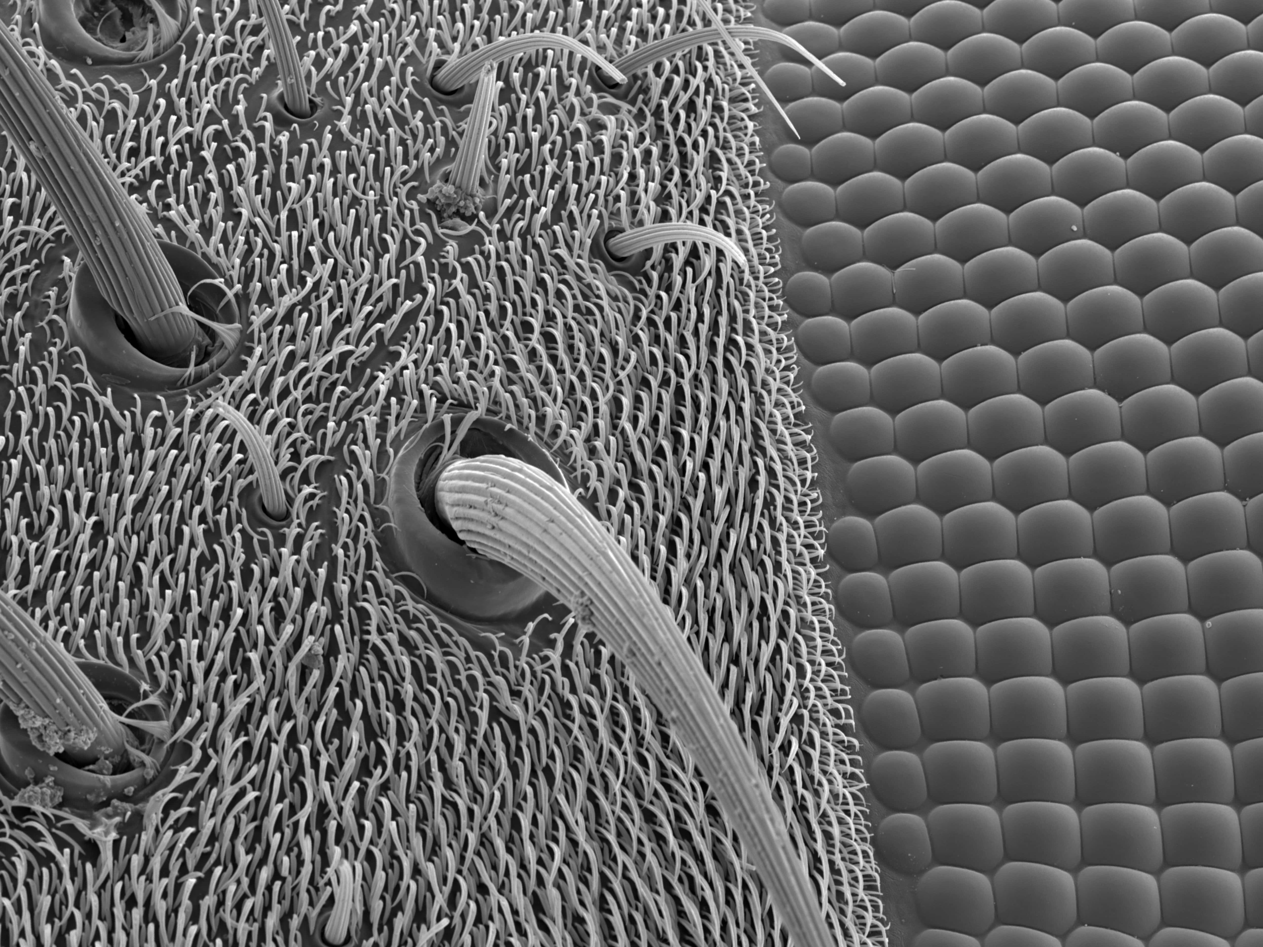
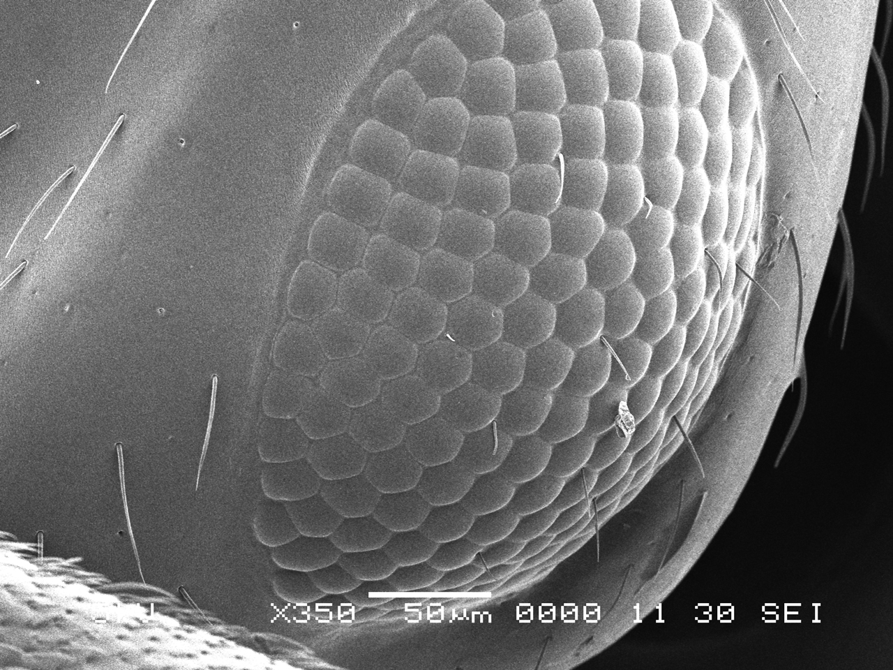
Head and compound eyes (Fly) | Amazing Nano World | JEOL

New Electron Microscope facility opened at University of Aberdeen | News |  The University of Aberdeen
New Electron Microscope facility opened at University of Aberdeen | News | The University of Aberdeen
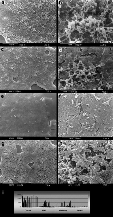
Scanning Electron Microscope Eye Structure Analysis

Scanning Electron Microscopy

Iris of the eye, SEM - Stock Image - P424/0106 - Science Photo Library

Impression cytology with scanning electron microscopy: a new method in the  study of conjunctival microvilli | Eye
Impression cytology with scanning electron microscopy: a new method in the study of conjunctival microvilli | Eye

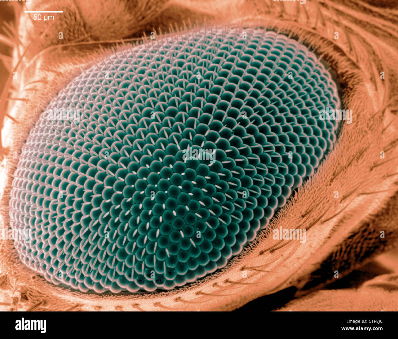
Scanning electron microscope image of an eye on a fruit fly Stock Photo -  Alamy
Scanning electron microscope image of an eye on a fruit fly Stock Photo - Alamy

Electron Microscopy — IKELOS